 96 382 55 77
A dental CBCT scan — short for Cone Beam Computed Tomography, also known as a 3D dental X-ray or dental cone beam CT — is a radiological imaging technique that produces a complete three-dimensional representation of the teeth, jaws, paranasal sinuses and facial bone structures, using a radiation dose far lower than that of a conventional medical CT scan.
If your dentist has told you that you need “a dental CT” or “a 3D scan” before getting implants, having a wisdom tooth removed or starting orthodontic treatment, this is exactly what they mean. Understanding what information it provides and why it is necessary in certain cases will help you approach your treatment with greater confidence.
At Asensio Dental Clinic, Dr. Lucía Asensio Romero (Registration No. 46002287) has the Planmeca PROMAX 3D system with integrated CBCT technology, allowing complete three-dimensional diagnosis to be carried out in-house without external referral. The first visit is completely free of charge.
What is a dental CBCT scan and how does it work
A dental CBCT scanner is a state-of-the-art X-ray unit that emits a cone-shaped beam of radiation that rotates around the patient’s head in a single rotation lasting between 10 and 20 seconds. During that rotation it captures hundreds of two-dimensional images from different angles, which specialist software automatically reconstructs into a high-resolution three-dimensional model.
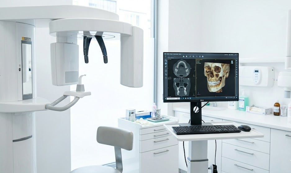
The result is a volume of data that the specialist can freely explore in all three planes of space — axial, coronal and sagittal — and from any angle, with the ability to isolate specific structures, measure distances to millimetre precision and plan treatments virtually before touching the patient.
The fundamental difference from a conventional panoramic X-ray — the OPG — is that the latter produces a flat two-dimensional image in which structures overlap and depth is lost. The CBCT adds the third dimension: it shows exactly where each structure is in space, how much bone is present at a specific point and what the relationship is between structures that would be superimposed in 2D.
The difference from a conventional medical CT is the radiation dose and the resolution in dental structures: CBCT delivers between 50% and 90% less radiation than a full-body CT and is specifically optimised for the resolution of hard tissues — bone and teeth — in the maxillofacial region, where a medical CT loses detail.
What is dental CBCT used for
CBCT is not a routine test carried out at every appointment: it is indicated in specific clinical situations where conventional two-dimensional imaging does not provide sufficient information to plan treatment safely. The main indications are as follows.
CBCT for dental implants
Planning dental implants is the most frequent indication for CBCT in dentistry. Before placing an implant it is essential to know the exact volume of available bone, bone density, the distance to the inferior alveolar nerve — whose injury would cause permanent paraesthesia — and proximity to the maxillary sinus. None of these measurements can be reliably obtained from a 2D panoramic X-ray.
With CBCT and 3D planning software, implants can be positioned virtually before surgery, a custom surgical guide can be designed to ensure the actual implant is placed exactly where planned, and the final prosthetic outcome can be anticipated in advance. In complex rehabilitations such as All-on-4 or zygomatic implants, CBCT is not optional — it is essential.
CBCT for wisdom tooth extraction
Extracting lower wisdom teeth carries a specific risk: proximity to the inferior alveolar nerve, whose injury can produce numbness or paraesthesia of the lower lip. When the panoramic X-ray suggests that the wisdom tooth roots may be in contact with the nerve canal, CBCT confirms whether there is a genuine relationship between the two structures or whether it is an artefact of the 2D image. This information determines whether the extraction can be safely carried out in the clinic or requires referral to an oral and maxillofacial surgeon.
CBCT in orthodontics
In orthodontics, CBCT is indicated for impacted teeth whose three-dimensional position cannot be determined in 2D, skeletal anomalies requiring surgical planning, airway analysis in patients with sleep apnoea, and evaluation of dental roots before and after orthodontic treatment to detect root resorption.
CBCT in endodontics and periapical pathology
In endodontics, CBCT allows detection of vertical root fractures not visible on conventional periapical X-rays, identification of untreated canals in retreatments, assessment of the true extent of periapical lesions and confirmation of bone healing after treatment. In oral medicine, CBCT is a first-line diagnostic tool for evaluating cysts, tumours and bone lesions of the jaws.
CBCT for TMJ and orofacial pain
TMJ evaluation with CBCT allows visualisation of the morphology of the mandibular condyle, detection of degenerative changes, erosions or condylar flattening, and assessment of symmetry between both joints. It is particularly useful in the differential diagnosis of orofacial pain when an articular origin is suspected or when conservative treatment has not produced the expected results.
| Indication | Why CBCT is needed | Key information provided |
|---|---|---|
| Dental implants | Precise measurement of available bone | Volume, bone density, distance to nerve and sinus |
| All-on-4 / zygomatic implants | Complex surgical planning | Virtual planning and surgical guide |
| Wisdom tooth extraction | Relationship with inferior alveolar nerve | 3D position of roots relative to the canal |
| Complex orthodontics / impacted teeth | Exact three-dimensional position | Eruption direction, relationship with adjacent roots |
| Endodontics / retreatments | Fractures and canals undetectable in 2D | Root fractures, canal anatomy |
| Bone pathology / cysts | True extent of the lesion | Borders, relationship with key structures |
| TMJ and orofacial pain | Mandibular condyle morphology | Degenerative changes, articular symmetry |
Radiation from dental CBCT: is it safe
Concern about radiation is completely understandable and has a data-based answer. The radiation dose from a dental CBCT varies according to the equipment, the field of view selected and the protocol used, but generally falls between 40 and 200 microsieverts (μSv) for a medium field of view — equivalent to between 3 and 15 conventional panoramic X-rays, and well below the 1,000–2,000 μSv of a conventional medical head CT.
To put this in perspective: the natural background cosmic radiation to which a person living in Europe is exposed amounts to approximately 2,400 μSv per year. A return transatlantic flight involves an exposure of between 50 and 100 μSv. A small-field dental CBCT — the type routinely used for single-unit implant planning — delivers around 40–60 μSv.
The Planmeca PROMAX 3D system used at Asensio Dental Clinic delivers an effective dose comparable to that of a conventional panoramic X-ray and up to 90% lower than a medical CT, thanks to low-dose protocols specifically optimised for dental diagnosis.
CBCT should always be clinically justified — it is not prescribed routinely but only when the information it provides is essential for treatment — and in groups with particular sensitivity such as children, pregnant women or patients with thyroid pathology, the lowest available dose protocols are applied.
What to expect on the day of your CBCT scan
The procedure is quick, painless and requires no preparation beforehand. The patient stands or sits inside the unit — in a position similar to that for a conventional panoramic X-ray, with no enclosed chamber and no sensation of claustrophobia — and remains still for the 10–20 seconds the X-ray arm takes to rotate. Removable dentures do not need to be removed unless specifically indicated by the specialist.
Images are obtained immediately and the specialist can begin evaluation straight away. In the implant planning workflow, the CBCT is integrated directly with the planning software to design the virtual implant positions and generate the surgical guide in the days that follow.
Dental CBCT in Valencia: where to get one
Not all dental clinics have their own CBCT equipment. In many cases the dentist prescribes the scan and the patient must attend an external diagnostic imaging centre, with the additional cost and delay this involves. At Asensio Dental Clinic the PROMAX 3D system with CBCT technology is available on-site, allowing diagnosis, planning and treatment to be carried out at a single centre with no waiting times or external referrals.
The CBCT is included in cases where it is clinically necessary within the diagnostic and treatment planning protocol for implantology, oral surgery and complex orthodontics. The first visit, which includes a full clinical examination, digital panoramic X-ray and intraoral scan, is completely free of charge.
Frequently asked questions about dental CBCT
Does a dental CBCT scan hurt?
No. The procedure is completely painless. The patient simply needs to remain still for the 10–20 seconds the image acquisition takes. There are no injections, no contrast agent and no physical contact with the equipment beyond the head positioning supports.
How much does a dental CBCT scan cost in Valencia?
The price of a dental CBCT in Valencia typically ranges between 80 and 200 euros depending on the field of view, the centre and whether the scan is carried out independently or as part of a treatment protocol. At Asensio Dental Clinic, CBCT is included in cases where it is necessary within the diagnosis and treatment plan, at no additional cost to the patient.
When is a CBCT necessary and when is a standard X-ray sufficient?
A conventional panoramic X-ray is sufficient for most routine check-ups, cavity diagnosis, general dental health evaluation and monitoring of straightforward treatments. CBCT is indicated when three-dimensional information is needed that 2D imaging cannot provide: implant planning, assessment of impacted teeth, diagnosis of root fractures, extent of bone lesions or detailed evaluation of the TMJ.
Can children have a dental CBCT scan?
Yes, with appropriate indications and using the lowest available dose protocols. In children and adolescents, CBCT is particularly indicated for the evaluation of retained permanent teeth, developmental anomalies and orthodontic planning in complex cases. Clinical justification must be especially rigorous in minors given that they are a group with greater sensitivity to radiation.
How often can a dental CBCT be taken?
There is no established frequency limit as long as each scan is clinically justified. In routine practice, a CBCT is prescribed when it is needed to plan a specific treatment — not periodically — and the cumulative dose over time is clinically negligible compared to natural background radiation.
Can dental CBCT detect oral cancer?
CBCT is highly effective at detecting lesions that affect bone — cysts, bone tumours, bone metastases — but it is not the appropriate test for diagnosing soft tissue lesions such as mucosal oral cancer, which requires direct clinical examination and biopsy. For early detection of oral cancer, regular clinical examination by a specialist in oral medicine remains the fundamental tool.